
Ficheru:DTI-sagittal-fibers.jpg
Tamañu d'esta previsualización: 643 × 600 pixels. Otres resoluciones: 257 × 240 pixels | 515 × 480 pixels | 1021 × 952 pixels.
Ficheru orixinal (1021 × 952 píxels, tamañu de ficheru: 294 kB, triba MIME: image/jpeg)
|
Resume
| Descripción |
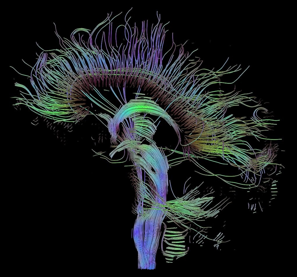
English: Visualization of a DTI measurement of a human brain. Depicted are reconstructed fiber tracts that run through the mid-sagittal plane. Especially prominent are the U-shaped fibers that connect the two hemispheres through the corpus callosum (the fibers come out of the image plane and consequently bend towards the top) and the fiber tracts that descend toward the spine (blue, within the image plane)
Français : Visualisation d'une mesure DTI d'un cerveau humain. Ce qui est représenté sont des faisceaux de fibres reconstruits qui traversent le plan demi-sagittal. On observe les fibres en U qui connectent les deux hémisphères à travers le corps calleux, qui sont particulièrement importantes (les fibres sortent du plan de l'image et par conséquent se courber vers le haut) ainsi que les faisceaux de fibres qui descendent vers la colonne vertébrale (bleu, dans le plan de l'image)
Deutsch: Traktographie-Verfahren rekonstruieren aus den Messdaten der Diffusions-Tensor-Bildgebung den anzunehmenden Verlauf größerer Nervenbahnen. Hier dargestellt sind die Ergebnisse für ein menschliches Gehirn; um die Übersichtlichkeit zu wahren, beschränkt sich die Abbildung auf Bahnen, die die Medianebene schneiden. Insbesondere sind dies die U-förmigen Faserbündel, die die beiden Hirnhälften verbinden (sie durchstoßen die Bildebene und sind nach oben gebogen) sowie die Faserbündel, die zum Rückenmark ziehen (blau dargestellt, liegen innerhalb der Bildebene) |
| Data | |
| Fonte | Trabayu propiu |
| Autor | Thomas Schultz |
| Permisu (Cómo reutilizar esti ficheru) |
Rendering is own work, using a modified version of the BioTensor application developed at the University of Utah. The dataset is courtesy of Gordon Kindlmann at the Scientific Computing and Imaging Institute, University of Utah, and Andrew Alexander, W.M. Keck Laboratory for Functional Brain Imaging and Behaviour, University of Wisconsin, Madison. It is publicly available from [1] |
Llicencia
Yo, el titular de los drechos d'autor d'esta obra, la espublizo baxo les siguientes llicencies:

|
Autorízase la copia, distribución y/o cambéu d'esti documentu baxo los términos de la Llicencia de documentación llibre GNU, versión 1.2 o cualesquier otra que nel futuru espublice la Free Software Foundation; ensin seiciones invariables, testos de portada, nin testos de contraportada. S'inclúi una copia de la llicencia na seición titulada GNU Free Documentation License. |
| Esti ficheru ta disponible baxo la llicencia Creative Commons Reconocimientu-Compartir igual 3.0 xenérica. | ||
| ||
| Esta etiqueta de llicencia s'amestó a esti ficheru como parte del anovamientu de la llicencia GFDL. |
Este archivo se encuentra bajo la licencia Creative Commons de Atribución/Compartir-Igual 2.5 Genérica, 2.0 Genérica y 1.0 Genérica.
- Ye llibre:
- pa compartir – pa copiar, distribuir y comunicar públicamente la obra
- pa remezclar – p'adautar la obra
- Baxo les condiciones siguientes:
- reconocimientu – Tienes de dar el créitu apropiáu, apurrir un enllaz a la llicencia ya indicar si realizasti dalgún cambéu. Puedes faelo de cualquier mou razonable ,pero non de manera que suxera l'encontu del autor pa ti o pal usu que faigas.
- compartir igual – Si entemeces, tresformes o te bases nesti material, tienes de distribuir les tos contribuciones baxo la mesma llicencia o una compatible cola orixinal.
Pues seleicionar la llicencia que prefieras.
Historial del ficheru
Calca nuna fecha/hora pa ver el ficheru como taba daquella.
| Data/Hora | Miniatura | Dimensiones | Usuariu | Comentariu | |
|---|---|---|---|---|---|
| actual | 10:42 13 och 2017 | | 1021 × 952 (294 kB) | Mikael Häggström | Minor crop of black areas at the top and bottom |
| 16:22 22 set 2006 |  | 1021 × 1125 (203 kB) | Thomas Schultz | {{Information |Description=Visualization of a DTI measurement of a human brain. Depicted are reconstructed fiber tracts that run through the mid-sagittal plane. Especially prominent are the U-shaped fibers that connect the two hemispheres through the corp |
Usu del ficheru
Nun hai páxines qu'usen esti ficheru.